Platelet-Rich Plasma: Healing Diabetic Ulcers Faster
How Your Blood Becomes Powerful Chronic Wound Medicine
Imagine if the most powerful medicine for your wound came from inside your own body. That is exactly what platelet-rich plasma (PRP) offers for people struggling with diabetic ulcers. This regenerative treatment concentrates natural healing proteins already present in your blood and delivers them directly to wounds that have refused to close for weeks or months. The results coming from the latest high-quality research are difficult to ignore.
Diabetes affects over 537 million people worldwide, and roughly one in four will develop a diabetic foot ulcer during their lifetime. These are not simple wounds. They are chronic, non-healing injuries caused by nerve damage, poor circulation and chronic inflammation working together to block normal repair. Without intervention, 20% of patients with diabetic foot ulcers eventually lose a limb. After amputation, mortality exceeds 70% within five years. These numbers make finding better treatments an urgent medical priority.
Traditional wound care involves cleaning, dressing changes and infection management. These approaches help, but they do not address the biological problems at the root of diabetic wound failure. This is where platelet-rich plasma enters as a genuine game changer, backed now by multiple large meta-analyses and network meta-analyses comparing it directly against every advanced therapy available. Before exploring what the science shows, it helps to understand what PRP actually is and how it works. If you are new to PRP, our overview article on PRP’s healing power covers the fundamentals in full detail.
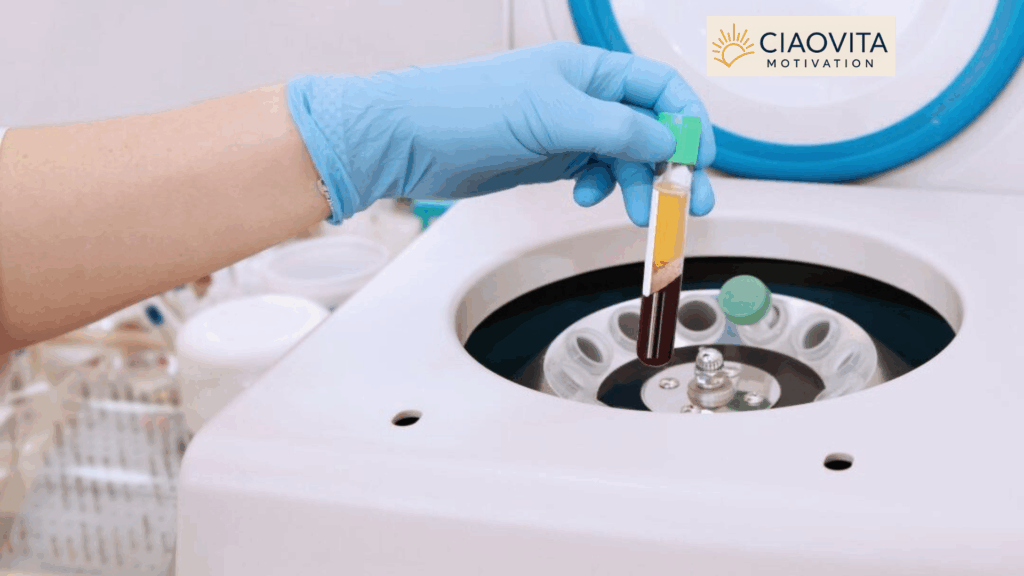
What Exactly Is Platelet-Rich Plasma?
Platelet-rich plasma starts with a routine blood draw. The sample then goes into a centrifuge — a machine that spins at high speed to separate blood components by density. The process creates three distinct layers: plasma at the top, a middle layer called the “buffy coat” containing platelets and white blood cells, and red blood cells at the bottom.
Platelets are tiny cells most people associate only with blood clotting. In reality, each platelet contains 50 to 80 alpha granules packed with hundreds of bioactive proteins, including powerful growth factors that coordinate tissue repair. When concentrated through centrifugation, the resulting PRP contains platelet levels far above normal blood values. This concentration transforms platelets from passive clotting agents into an active healing toolkit.
The preparation process matters significantly. Centrifuge speed, spinning time, the number of centrifugation rounds, and the anticoagulant used during collection all influence the final product. Some practitioners add a second spin to concentrate platelets further. Others use commercial automated systems. Research suggests an optimal platelet concentration of around 1.0 to 1.5 million platelets per microliter — concentrations below this threshold may not deliver enough growth factors, while very high concentrations may paradoxically reduce effectiveness through receptor downregulation.
Some preparations include an activation step using thrombin or calcium chloride, which triggers immediate growth factor release and creates a gel-like consistency ideal for topical wound application. Others skip activation, allowing gradual release after the PRP contacts wound tissue. Neither approach has proven definitively superior, and both show consistent benefits across clinical studies.
The key growth factors released by platelet-rich plasma include platelet-derived growth factor (PDGF), which recruits repair cells to the wound site; vascular endothelial growth factor (VEGF), which promotes new blood vessel formation; transforming growth factor-beta (TGF-β), which stimulates collagen production; fibroblast growth factor (FGF), which supports cell multiplication; and epidermal growth factor (EGF), which drives new skin formation. These molecules work as a coordinated team, each amplifying the others’ effects.
Understanding how each growth factor contributes explains why PRP has advantages over therapies that deliver only a single factor. You can explore this comparison in depth in our article on PRP for joint pain and sports injuries, which discusses how the same growth factor cocktail restores damaged tissue in completely different contexts.
How Platelet-Rich Plasma Promotes Wound Healing
Normal wound healing follows four overlapping phases: hemostasis (stopping bleeding), inflammation, proliferation (new tissue formation) and remodeling. Diabetic ulcers get trapped, typically in the inflammatory phase. Instead of progressing toward repair, the wound environment stays chronically inflamed. High glucose levels, oxidative stress and mitochondrial dysfunction damage blood vessels and nerve endings, creating conditions that actively resist healing. Understanding this failure point is essential to understanding why platelet-rich plasma works so well for diabetic wounds.
PRP addresses multiple problems simultaneously. When applied to a wound, it floods the healing environment with the full spectrum of growth factors described above. PDGF attracts fibroblasts — the cells that produce structural proteins — directly to the wound site. VEGF stimulates the formation of new capillaries, restoring the blood supply that diabetes has compromised. TGF-β drives collagen synthesis, building the structural scaffolding that new tissue needs to organize itself.
Beyond growth factors, PRP contains proteins that actively resolve excessive inflammation. One remarkable interaction involves platelets and neutrophils — a type of white blood cell that normally produces inflammatory molecules called leukotrienes. When platelets come into contact with neutrophils, they convert these inflammatory substances into lipoxins, powerful anti-inflammatory compounds that help end the inflammatory cycle rather than perpetuate it. This biological switch is one of the reasons PRP helps wounds exit the chronic inflammatory state where diabetic ulcers are trapped.
The treatment also demonstrates antimicrobial effects. Diabetic ulcers frequently become infected, and infection dramatically worsens outcomes. Multiple studies report lower infection rates in PRP-treated wounds compared to standard dressings, though the precise mechanisms require further investigation. This combination of pro-healing and anti-infective properties makes PRP uniquely well-suited to the diabetic wound environment. For a broader perspective on how diabetes impairs normal tissue healing, our article on diabetes and orthopedic surgery risks provides important context on how elevated blood sugar disrupts biological repair across multiple tissue types.
💡 KEY MECHANISM — Why PRP Works in Diabetic Wounds Diabetic ulcers get stuck in chronic inflammation because high blood sugar, nerve damage and poor circulation block the normal healing sequence. Platelet-rich plasma provides a concentrated delivery of growth factors (PDGF, VEGF, TGF-β, FGF, EGF) that restart the stalled repair process, resolve excess inflammation, promote new blood vessel formation and support collagen production — all simultaneously, from the patient’s own blood. |
What the Research Really Shows
The evidence base for platelet-rich plasma in diabetic foot ulcer treatment has grown substantially in recent years, driven by multiple large systematic reviews and meta-analyses that pool data across dozens of randomized controlled trials.
The most comprehensive evidence comes from a 2023 systematic review and meta-analysis published in the Journal of Wound Care (Su et al.). This study analyzed 17 randomized controlled trials involving 1,303 participants — 649 receiving autologous PRP and 654 receiving standard care. It confirmed that PRP significantly improves complete healing rates and reduces time to healing, with no increase in adverse events. The primary outcome — proportion of complete ulcer healing — favored PRP consistently across study populations.
A second 2023 meta-analysis published in the Journal of Orthopaedic Surgery and Research (Deng et al.) analyzed 22 randomized controlled trials and produced some of the most precise quantitative data available:
- Healing rate: RR = 1.42 (95% CI: 1.30–1.56, p < 0.001) — 42% better than standard care
- Healing time reduction: MD = –3.13 days (95% CI: –5.86 to –0.39, p < 0.001)
- Ulcer area reduction: MD = 1.02 (95% CI: 0.51–1.53, p < 0.001)
- Amputation rate: RR = 0.35 (95% CI: 0.15–0.83, p < 0.001) — 65% reduction in amputation risk
- Adverse events: RR = 0.96 (p > 0.05) — no increase compared to conventional therapy
That fourth finding deserves emphasis. A 65% reduction in amputation risk means that for every three amputations that would occur with standard care, PRP potentially prevents two of them. Given that amputation in this patient group carries a five-year mortality rate exceeding 70%, this outcome carries enormous clinical significance. For context on how tissue regeneration approaches are reshaping multiple areas of medicine, see our article on stem cell therapy and natural healing.
A 2023 systematic review published in Frontiers in Endocrinology (OuYang et al.) went beyond standard efficacy measures to evaluate outcomes that matter directly to patients — ulcer recurrence, hospitalization costs and quality of life scores. This study analyzed 20 controlled trials and found PRP reduces recurrence rates in addition to improving initial healing. It also confirmed that combining PRP with other modalities amplifies these benefits.
Comparing PRP to Other Advanced Therapies
One of the most important questions in diabetic wound care is not just “does PRP work?” but “how does PRP compare to everything else available?” A landmark 2024 network meta-analysis published in Frontiers in Endocrinology (OuYang et al.) answered this question directly by analyzing 57 randomized controlled trials involving 4,826 patients across 11 different treatment categories.
The 11 treatments compared were: platelet-rich plasma (PRP), negative pressure wound therapy (NPWT), hyperbaric oxygen therapy (HBOT), topical oxygen therapy (TOT), ultrasonic debridement (UD), acellular dermal matrix (ADM), stem cells (SCs), and various combinations. Each was measured against the current standard of care (SOC) on five outcomes: complete healing rate, ulcer area reduction, healing time, amputation rate and adverse events.
The results placed PRP among the top-ranking individual treatments on multiple outcomes:
- Complete healing rate: PRP, HBOT, TOT, ADM and stem cells all significantly outperformed standard care in both direct and network analyses.
- Healing time: Both PRP and NPWT significantly shortened healing time versus standard care.
- Amputation rate: PRP effectively reduced amputation rates — the only individual therapy to do so with statistical significance in this analysis.
- Adverse events: PRP significantly reduced adverse event incidence compared to standard care.
- Combination therapy: PRP combined with NPWT outperformed NPWT alone for complete healing. The PRP + UD + NPWT combination produced the shortest healing times of all interventions tested.
The network meta-analysis’s ranking system placed combined PRP approaches consistently at the top. The SOC group ranked last across every outcome category. This finding carries a clear message: standard wound care alone is no longer adequate for complex diabetic foot ulcers when evidence-based alternatives exist.
A 2025 updated network meta-analysis published in Frontiers in Endocrinology (Tian et al.) analyzed 51 randomized controlled trials with 3,401 patients and focused on comparing PRP against other individual growth factor therapies — including epidermal growth factor (EGF), platelet-derived growth factor (PDGF), fibroblast growth factor (FGF), vascular endothelial growth factor (VEGF) and granulocyte colony-stimulating factor (G-CSF). The analysis confirmed that PRP improves healing rate and shortens healing time versus standard care. Importantly, PRP was associated with the most favorable safety profile among all growth factor therapies — ranking first for adverse event reduction and amputation prevention. EGF ranked slightly higher for healing rate alone, but PRP’s combination of efficacy and safety made it the most clinically balanced option overall.
This safety advantage makes practical sense. Because PRP is autologous — drawn from the patient’s own blood — the body recognizes its components rather than treating them as foreign substances. Allergic reactions are extremely rare. The process involves no synthetic compounds, no recombinant proteins and no animal-derived materials. Understanding why metabolic conditions like poorly controlled diabetes impair normal healing is essential for patients and families. Our article on metabolic syndrome risks and solutions explains the metabolic foundations that make wound healing so challenging in diabetic patients.
📊 RESEARCH SUMMARY — Key Numbers You Should Know • 1,303 patients across 17 RCTs: autologous PRP improves complete healing (Su et al., J Wound Care 2023) • 42% better healing rate (RR = 1.42) and 65% lower amputation risk (RR = 0.35) (Deng et al., 2023) • 4,826 patients across 57 RCTs: PRP ranks top for healing AND safety vs. 11 different advanced therapies (OuYang et al., 2024) • PRP + NPWT combination: superior to both treatments used alone (NMA 2024) • Best safety profile among all growth factor therapies tested (Tian et al., 2025) |
Practical Considerations and What Patients Should Know
Despite impressive evidence, several practical factors affect how platelet-rich plasma therapy reaches patients with diabetic foot ulcers. Understanding these factors helps set realistic expectations and guides productive conversations with healthcare providers.
Availability and regulation vary significantly by country. In the United States, PRP has greater clinical access, though insurance coverage is inconsistent. In Brazil and many European countries, regulatory agencies currently classify PRP for wound healing on an experimental or conditional basis, meaning access depends on individual institutions and specialist centers. This situation is evolving rapidly as the evidence base strengthens — what was experimental five years ago increasingly appears in treatment guidelines today.
Cost presents another real barrier. While PRP preparation is relatively straightforward compared to therapies like hyperbaric oxygen chambers or engineered skin substitutes, it requires centrifuge equipment and trained personnel. Commercial automated PRP systems simplify preparation and improve consistency, but add expense. For patients already managing the substantial costs of diabetes care — medications, monitoring equipment, specialist visits — out-of-pocket PRP expenses may be prohibitive without insurance support.
Standardization remains a significant challenge across the entire field. Blood collection protocols, anticoagulant choices, centrifuge parameters, activation methods and application frequency all vary between practitioners and research centers. This inconsistency creates real-world variability in outcomes and makes it difficult to replicate the best results from clinical trials in community settings. Professional organizations in wound care and regenerative medicine are actively working toward consensus protocols, but standardization has not yet been fully achieved.
Patient selection matters considerably. Most clinical trials excluded people with platelet disorders, severe uncontrolled infections, malignancy, platelet counts below certain thresholds or certain immunosuppressive medications. These exclusions make clinical sense for research validity, but they mean that real-world populations — who often have multiple comorbidities — may respond differently from trial participants. More research is needed to refine selection criteria and identify which patient profiles benefit most.
The foundation of effective treatment always remains comprehensive diabetic wound care. Platelet-rich plasma works best when built on solid fundamentals: optimal glucose control, aggressive infection management, appropriate debridement of dead tissue, offloading pressure from the wound, and regular professional monitoring. The 2024 network meta-analysis confirmed that combinations of advanced therapies consistently outperform single treatments — reinforcing that PRP is a powerful addition to, not a replacement for, complete diabetic care.
Questions worth raising with your healthcare provider include:
- Is PRP available at your facility or a nearby specialist center?
- Does your specific wound type and severity qualify based on current clinical guidelines?
- What application protocol does the center use — topical gel, direct injection or a combination?
- How does the cost compare to other available advanced therapies, and is any coverage possible?
- How will results be monitored, and at what point would the protocol be adjusted?
Looking forward, researchers are working to identify biomarkers that predict which patients will respond best to PRP — moving toward a personalized medicine model where treatment protocols match individual biological characteristics. Scientists are also investigating optimal platelet concentrations, combination protocols with stem cells and engineered skin substitutes, and the specific dose-response relationships for each growth factor. The combination of PRP with stem cell therapy represents one of the most promising frontiers, with early results suggesting synergistic benefits that neither therapy achieves alone.
Conclusion: What the Evidence Means for You
The evidence supporting platelet-rich plasma for diabetic foot ulcers has moved well beyond preliminary. Multiple large meta-analyses — including a 2024 network meta-analysis comparing 11 therapies in 4,826 patients — consistently confirm that PRP improves complete healing rates by approximately 42%, reduces healing time, lowers amputation risk by 65% and maintains a safety profile that is equal to or better than all comparators. When combined with negative pressure wound therapy, outcomes improve further.
For the millions of people living with diabetes who develop a chronic wound, this represents genuine, evidence-based hope. Platelet-rich plasma harnesses the healing intelligence already present in your own blood, concentrating it precisely where the body’s repair mechanisms have failed. As protocols become more standardized and access improves, PRP is well-positioned to transition from advanced adjunct therapy to a standard component of diabetic wound care.
If you or a family member is managing a diabetic foot ulcer that is not responding to standard treatment, the research now supports a direct conversation with your specialist about PRP. Combined with rigorous glucose control, appropriate offloading, infection management and close monitoring, platelet-rich plasma offers a scientifically validated path toward healing — and toward preserving the limb function that makes independent living possible.
References
1. Su YN, Li J, Feng DH, Lu RR, Dong GX, Zhao DY. Efficacy and safety of autologous platelet-rich plasma for diabetic foot ulcers: a systematic review and meta-analysis. J Wound Care. 2023;32(12):773-786. https://pubmed.ncbi.nlm.nih.gov/38060413/
2. Deng J, Yang M, Zhang X, Zhang H. Efficacy and safety of autologous platelet-rich plasma for diabetic foot ulcer healing: a systematic review and meta-analysis of randomized controlled trials. J Orthop Surg Res. 2023;18(1):370. https://pubmed.ncbi.nlm.nih.gov/37202812/
3. OuYang H, Tang Y, Yang F, Ren X, Yang J, Cao H, Yin Y. Platelet-rich plasma for the treatment of diabetic foot ulcer: a systematic review. Front Endocrinol. 2023;14:1256081. https://pubmed.ncbi.nlm.nih.gov/38169990/
4. OuYang H, Yang J, Wan H, Huang J, Yin Y. Effects of different treatment measures on the efficacy of diabetic foot ulcers: a network meta-analysis. Front Endocrinol. 2024;15:1452192. https://pubmed.ncbi.nlm.nih.gov/39377075/
5. Tian J, Yao G, Tian T, Li X, Li S, Wu C, Zhang S. Comparison of the efficacy and safety of different growth factors in the treatment of diabetic foot ulcers: an updated network meta-analysis. Front Endocrinol. 2025. https://www.frontiersin.org/journals/endocrinology/articles/10.3389/fendo.2025.1614597/full
© 2025 Alice & Marcus Guimarães. All rights reserved.This site is proudly created with WordPress.